 ESPCI web site


Olivia du Roure
Biological cells sense and respond to mechanical stimuli. Central to this process is the actin cytoskeleton, a dynamic network of filaments. The assembly of this network is also responsible for cell locomotion in embryogenesis, wound healing and cancer metastasis.
Our team develops new experiments to elucidate the biophysical processes at play in cell mechanics and motility. We measure the mechanical properties and the force generation of reconstituted actin networks, whole cells and organelles.
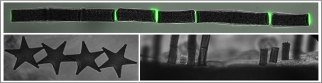
The general framework is to fabricate and use (super-para-) magnetic colloids, cylinders and micro-patterns to exert and measure forces on micro-objects. We use the dipolar attraction between two magnetic structures which develops nanoNewton forces which are relevant for biology. We take advantage of the self-organization properties of these system to multiply the number of measures and are thus able to characterize 100 to 1000 more structures than possible with other techniques.
Our current works and projects address different questions related to cell mechanics through experiments carried out with this approach:
Assembly and mechanics of growing actin networks.
We follow the growth of actin networks assembled in vitro from the surface of magnetic cylinders while subject to a load force. We link the viscoelastic properties of these network to their growth behaviour (PhD Pierre Bauër 2012-2015, Coll Martin Lenz, LPTMS, Orsay)
Mechanics of yeast actin networks.
We use genetically modified yeast extract to probe the role of different proteins into the elastic and plastic properties of actin gels through a top-down approach (Coll. Alphée Michelot IBDM, Marseille. PhD Jessica Planade (2013-2016))
Mechanotransduction of actin networks.
We investigate how the biochemical binding and unbinding of actin’s partners are influenced by the bending and stretching of actin filaments in a network (Coll G Romet-Lemonne, IJM, Paris. ANR Muscactin 2016-2020)
Transmigration through epithelial cells.
We use epithelial cells grown on magnetic micro-patterns to measure the spatiotemporal mechanical response of cells in a layer to a stimuli, that can explain the migration of white blood cells out of blood vessels. (Coll. Julien Husson, LadhyX, Palaiseau).
Intracellular response to forces.
We use magnetic cylinders ingested by dendritic cells to exert forces inside the cell to understand how the cytoskeleton and the nucleus change during locomotion (Collab. Matthieu Piel, Inst. Curie, Paris. PhD Valentin Laplaud (2016-2019))